

Case Example 1:
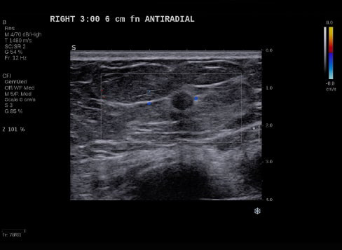
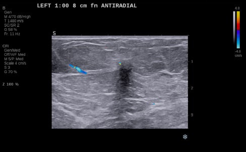
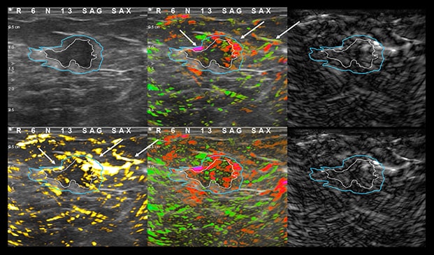
77-year-old female recalled for diagnostic imaging on new 0.7 cm mass seen on right breast. Ultrasound imaging demonstrates a round mass with an echogenic rim and minimal doppler signal.
Case Example 1: Observations
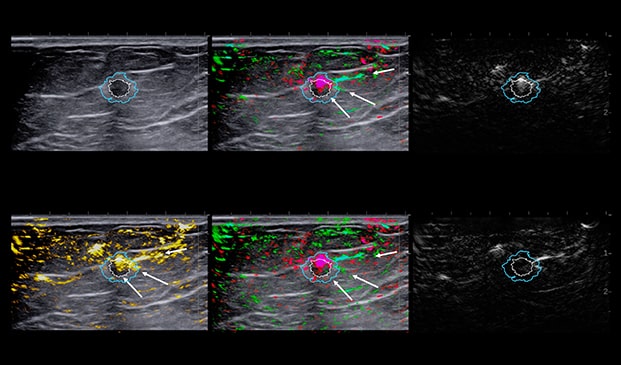
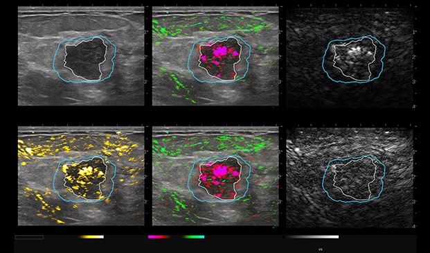
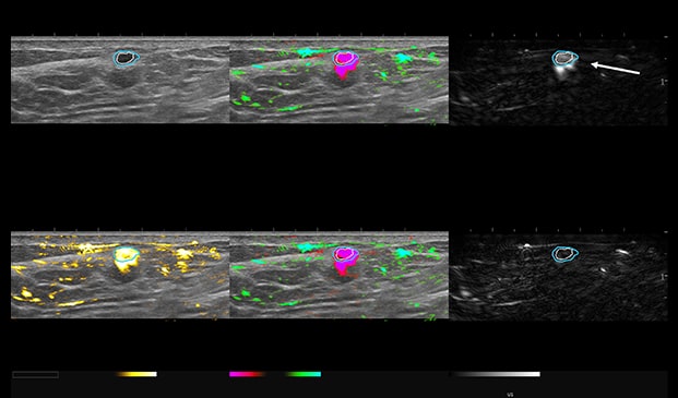
With OA, we can see deoxygenated hemoglobin both internally to the mass and externally in the boundary and periphery of the mass as an intense pink blush as it invades normal breast tissues.
On the combined map (upper middle) and relative map (lower middle), and even in the total hemoglobin map (lower left), we can see perpendicular vessels in the peripheral zone that enter through the boundary zone. These are characteristics seen in cancerous masses.
Invasive Breast Carcinoma Grade II – with Lobular Feature
- ER+
- HER2-
- PR+
- Ki67=15%
This patient was up-classified to a BI-RADS 5. Luminal B masses typically present with both internal and external OA features.
Case Example 2:
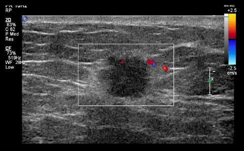
48-year-old female, presented with an abnormal mammogram/tomo. 1.9 cm mass seen on ultrasound, negative doppler.
Case Example 2: Observations
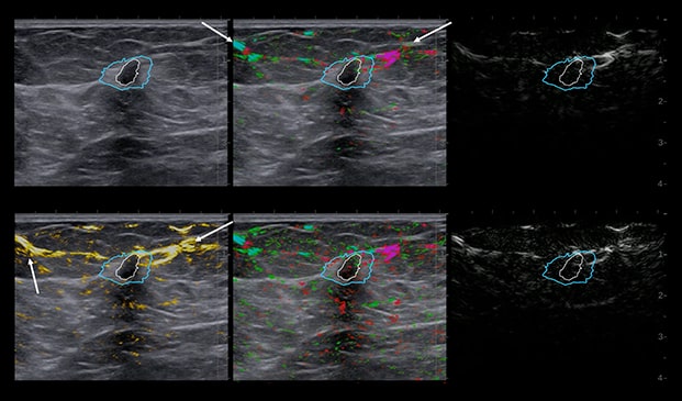
Deoxygenated hemoglobin internal to the mass is represented by intense disorganized or polymorphic pink colorization seen on the 6-up image.
These are typical malignant characteristics on OA which are represented on the combined map (upper middle), total hemoglobin (lower left) and relative map (lower middle).
Invasive Ductal Carcinoma Grade III
- ER-
- HER2-
- PR-
- Ki67=80%
The Mass was up-classified to BI-RADS 4C. Biopsy confirmed triple negative cancer. TNC masses typically present with intense deoxygenated mainly internal vessels. Masses with high Ki67 tend to have a lot of internal vessels mainly in the internal and boundary zones, unless there is necrosis.
Case Example 3:
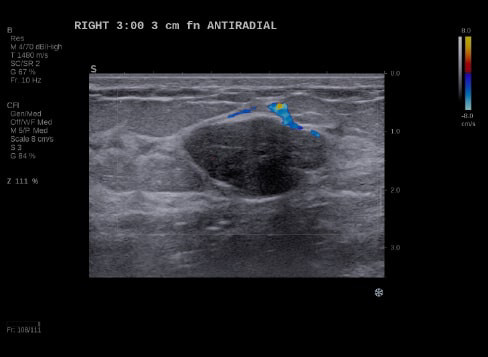
24-year-old female presents with a 2.7 cm palpable mass in her right breast. Ultrasound imaging demonstrates a solid mass with negative color doppler.
Case Example 3: Observations
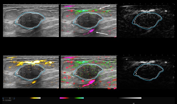
The mass does not reveal any suspicious internal signal on the combined map with paired draping vessels anteriorly and a deoxygenated vessel posteriorly. Draping vessels are typical benign characteristics when seen in OA, as the mass pushes the vessels away versus penetrating or feeding the mass in a cancer OA image.
Balanced mixture of well-organized red and green vessels, small in size with large periphery vessels as seen in the OA Relative map.
- No biopsy - LOM BI-RADS 2
- Fibroadenoma
Mass down-classified from BI-RADS 3 to BI-RADS 2. Typical OA characteristics seen in fibroadenomas.
Case Example 4:
53-year-old female, called back for diagnostic imaging. A 5 mm mass seen on ultrasound with complex, negative doppler signal.
Case Example 4: Observations
OA imaging demonstrates an increase in an intense pink colorization with an “eggshell sign” seen on the short wavelength. The raw short wavelength image represents deoxygenated hemoglobin and the long wavelength map in the lower right represents the raw oxygenated Hgb from the two lasers.
The eggshell pattern on the short wavelength is a benign feature, in addition to the intense pink colorization appearing as a uniform bloom versus a polymorphic (disorganized) pattern often seen with malignant masses.
Angiogenesis can be malignant or benign. Here we see physiologic angiogenesis associated with acute inflammation.
- Fibrocystic, Proliferative
- Inflamed Cyst
This patient was biopsied, and results were fibrocystic inflamed cyst. Mass down-classified from BI-RADS 4A to BI-RADS 2.
Case Example 5:
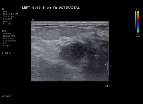
59-year-old female presents with a new mammographic asymmetry on the left breast.
A 1.2 cm mass is seen on ultrasound with some doppler signal along periphery of mass.
Case Example 5: Observations
With OA imaging we can see negative internal findings but positive external features in the boundary and peripheral zones.
We can see feeding and draining vessels or “whiskers” in the boundary and peripheral zones on OA.
Radiating vessels continue from the peripheral zone into the boundary zone and count as features in both zones.
Invasive Ductal Carcinoma Grade I
- ER+
- HER2-
- PR-
- Ki67=10%
The mass was up-classified to BI-RADS 5. Unlike TNC, luminal A masses typically present with external features in the boundary and peripheral zones and no OA internal features. Typically, luminal A masses have a low Ki67 and tend to have less internal angiogenesis and deoxygenation.
Case Example 6:
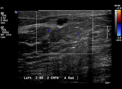
58-year-old female presents with a new mammographic finding. On ultrasound, we can see a 1.2 cm mass with minimal Doppler signal internally and just outside the mass in the boundary zone.
Case Example 6: Observations
In the OA imaging we can see an increase in deoxygenated hemoglobin both internally to the mass and in the boundary zone of the mass as seen on the relative and total hemoglobin OA maps.
Invasive Ductal Carcinoma Grade III
- ER-
- HER2+
- PR-
- Ki67=N/A
This mass was biopsied and was a grade III invasive ductal carcinoma, HER2 positive molecular subtype breast cancer.