
Imagio® delivers diagnostic confidence at the point of care
With new technology that has never been available before, Imagio® provides substantially improved confidence in breast cancer diagnostics using a non-invasive, real-time OA/US scan, supported by our proprietary AI-driven SenoGram® decision support. The simplified and streamlined care pathway offers the first FDA-approved device to effectively reduce the delays, discomfort, and costs of diagnosing breast cancer.
Radiologists Patient Experience Practice Efficiency Read our eBook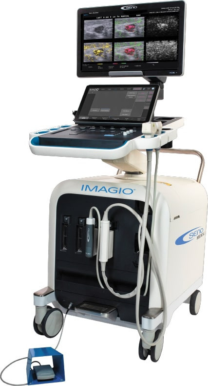
Laser light in. Sound out.
The new imaging modality to diagnose breast cancer in real-time
By transmitting laser light through suspicious breast tissue, Imagio® returns ultrasound signals that can display both functional and anatomical information about the relative blood and oxygen concentration. This enables clinicians to differentiate malignant and benign tissue: in a full-color display.
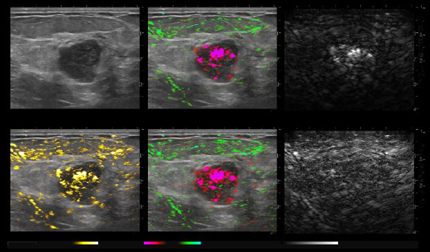
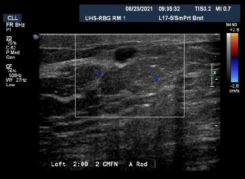
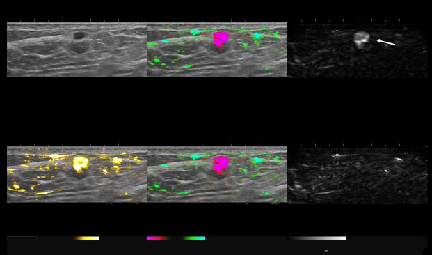
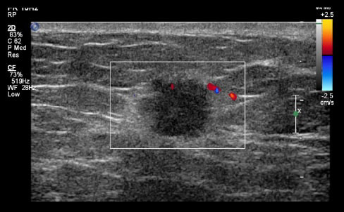
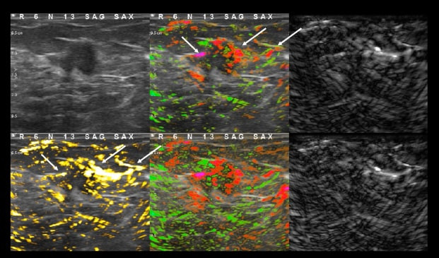
77-year-old female recalled for diagnostic imaging on new 0.7 cm mass seen on right breast. Ultrasound imaging demonstrates a round mass with an echogenic rim and minimal doppler signal.
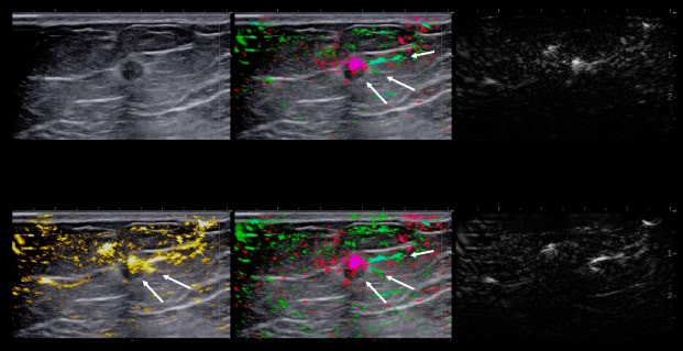
With OA, we can see deoxygenated hemoglobin both internally to the mass and externally in the boundary and periphery of the mass as an intense pink blush as it invades normal breast tissues.
On the combined map (upper middle) and relative map (lower middle), and even in the total hemoglobin map (lower left), we can see perpendicular vessels in the peripheral zone that enter through the boundary zone. These are characteristics seen in cancerous masses.
Invasive Breast Carcinoma Grade II – with Lobular Feature
- ER+
- HER2-
- PR+
- Ki67=15%
This patient was up-classified to a BI-RADS 5. Luminal B masses typically present with both internal and external OA features.
48-year-old female, presented with an abnormal mammogram/tomo. 1.9 cm mass seen on ultrasound, negative doppler.
Deoxygenated hemoglobin internal to the mass is represented by intense disorganized or polymorphic pink colorization seen on the 6-up image.
These are typical malignant characteristics on OA which are represented on the combined map (upper middle), total hemoglobin (lower left) and relative map (lower middle).
Invasive Ductal Carcinoma Grade III
- ER-
- HER2-
- PR-
- Ki67=80%
The Mass was up-classified to BI-RADS 4C. Biopsy confirmed triple negative cancer. TNC masses typically present with intense deoxygenated mainly internal vessels. Masses with high Ki67 tend to have a lot of internal vessels mainly in the internal and boundary zones, unless there is necrosis.
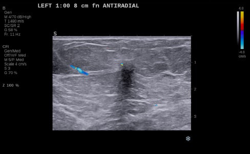
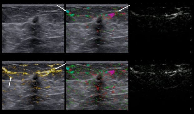
Positive lymph nodes were also imaged in this patient’s axilla. Within the thickened cortex of the lymph node, we can see an increase in feeding and draining vessels containing deoxygenated blood. Note the anterior extra hilar transcapsular vessels located anteriorly.
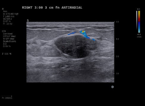
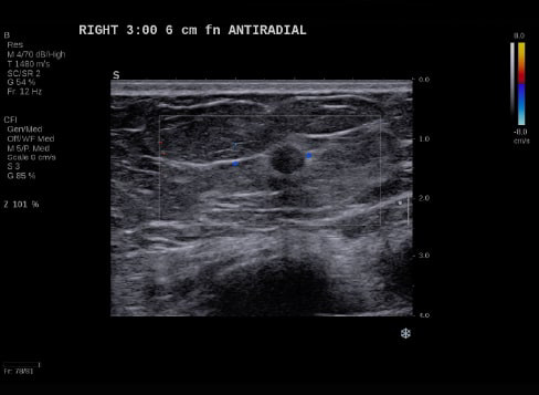
24-year-old female presents with a 2.7 cm palpable mass in her right breast. Ultrasound imaging demonstrates a solid mass with negative color doppler.
The mass does not reveal any suspicious internal signal on the combined map with paired draping vessels anteriorly and a deoxygenated vessel posteriorly. Draping vessels are typical benign characteristics when seen in OA, as the mass pushes the vessels away versus penetrating or feeding the mass in a cancer OA image.
Balanced mixture of well-organized red and green vessels, small in size with large periphery vessels as seen in the OA Relative map.
- No biopsy - LOM BI-RADS 2
- Fibroadenoma
Mass down-classified from BI-RADS 3 to BI-RADS 2. Typical OA characteristics seen in fibroadenomas.
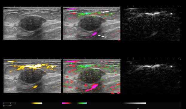
53-year-old female, called back for diagnostic imaging. A 5 mm mass seen on ultrasound with complex, negative doppler signal.
OA imaging demonstrates an increase in an intense pink colorization with an “eggshell sign” seen on the short wavelength. The raw short wavelength image represents deoxygenated hemoglobin and the long wavelength map in the lower right represents the raw oxygenated Hgb from the two lasers.
The eggshell pattern on the short wavelength is a benign feature, in addition to the intense pink colorization appearing as a uniform bloom versus a polymorphic (disorganized) pattern often seen with malignant masses.
Angiogenesis can be malignant or benign. Here we see physiologic angiogenesis associated with acute inflammation.
- Fibrocystic, Proliferative
- Inflamed Cyst
This patient was biopsied, and results were fibrocystic inflamed cyst. Mass down-classified from BI-RADS 4A to BI-RADS 2.
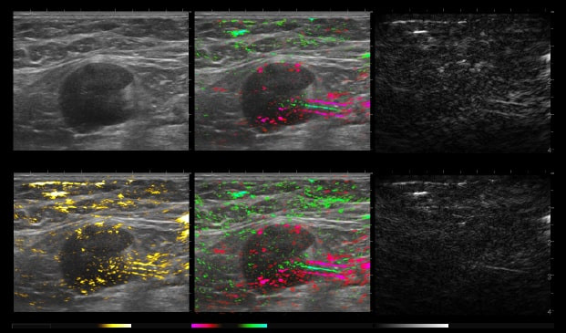
59-year-old female presents with a new mammographic asymmetry on the left breast.
A 1.2 cm mass is seen on ultrasound with some doppler signal along periphery of mass.
With OA imaging we can see negative internal findings but positive external features in the boundary and peripheral zones.
We can see feeding and draining vessels or “whiskers” in the boundary and peripheral zones on OA.
Radiating vessels continue from the peripheral zone into the boundary zone and count as features in both zones.
Invasive Ductal Carcinoma Grade I
- ER+
- HER2-
- PR-
- Ki67=10%
The mass was up-classified to BI-RADS 5. Unlike TNC, luminal A masses typically present with external features in the boundary and peripheral zones and no OA internal features.
Typically, luminal A masses have a low Ki67 and tend to have less internal angiogenesis and deoxygenation.
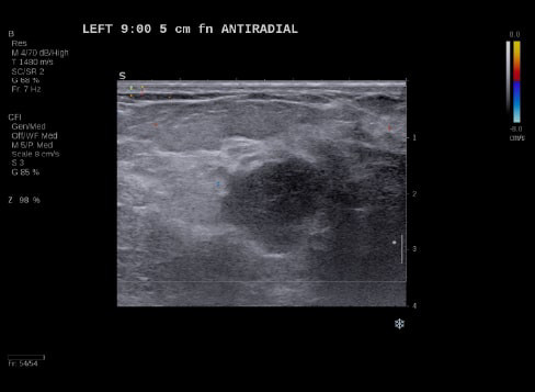
58-year-old female presents with a new mammographic finding. On ultrasound, we can see a 1.2 cm mass with minimal Doppler signal internally and just outside the mass in the boundary zone.
In the OA imaging we can see an increase in deoxygenated hemoglobin both internally to the mass and in the boundary zone of the mass as seen on the relative and total hemoglobin OA maps.
Invasive Ductal Carcinoma Grade III
- ER-
- HER2+
- PR-
- Ki67=N/A
This mass was biopsied and was a grade III invasive ductal carcinoma, HER2 positive molecular subtype breast cancer.
“Optoacoustic imaging quantifies additional functional information on tumor biology,… “paralleling” the dynamic data obtained with breast MRI, contrast-enhanced mammography, and molecular breast imaging, though without the cost of magnet time, ionizing radiation, or risk of allergic reactions from gadolinium-based or iodinated contrast material.“1
- Panigrahi B.

“This is game-changing for breast cancer imaging in that we get functional information—that we would normally get from advanced imaging—but with ultrasound.”
- Basak Dogan, MD
Director of Research at UT Southwestern Medical Center’s Harold C.Simmons Comprehensive Cancer Center

“This makes you a better radiologist, having more and deeper information. We can decide more certainly with this anatomic and functional information what is the right path for the patient.”
- Jeroen Veltman, MD, PhD
Assistant Professor, Chair, Dutch College of Breast Imaging (DCBI)

“Especially in large markets and large cities, where hospital systems compete against each other, it's nice to be able to say that our radiology practice offers the newset technology, including cutting-edge opto-acoustic imaging. When it comes to healthcare, women seek out technologies that can help them. This could be one.”
- Kenneth Kist, MD
Retired Radiologist at UT Health San Antonio

“The training was excellent, getting us comfortable with a new way of scanning. Once you scan a few patients, say five or six, you get the feel of the system and gain confidence in what you are seeing in the images.”
- Thanh Van, MD
Director of Breast Imaging and Intervention at UT Health San Antonio
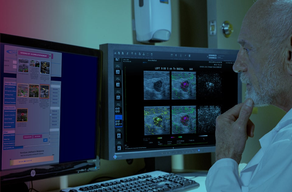
AI-based decision support
Accurately assign diagnostic BI-RADS
Proven to make clinicians more accurate, our proprietary SenoGram® provides AI-driven decision support in assessing likelihood of malignancy (LOM).
View SenoGram® brochure Interact with SenoGram®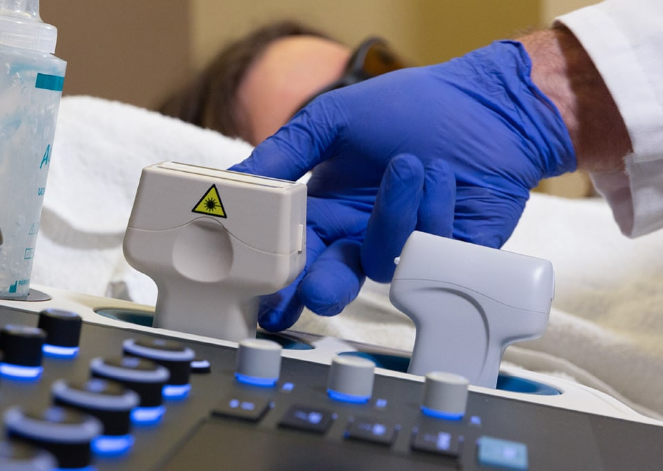
A simple transition that improves practice efficiency and profitability
Imagio® integrates into your existing diagnostic workflow over time, similar to how tomosynthesis was introduced. The technology can be easily adopted with the support of SenoGram® and training from a leading Breast Ultrasound expert, Dr. Tom Stavros through Seno University.
Request demo See ROI example
Our nationwide roadshow can bring Imagio® to your facility or you can start the conversation with a Seno rep today to get more details.
References
- Panigrahi B, Editorial Comment: The Value of Decision Support in Breast Ultrasound. AJR 2022 Dec 21
- Seiler SJ, Neuschler EI, Butler RS, Lavin PT, Dogan BE. Optoacoustic imaging with decision support for differentiation of benign and malignant breast masses: A 15-reader retrospective study. AJR Am J Roentgenol. December 7, 2022.