
How Imagio® works
Opto-acoustic (also called photoacoustic) imaging combines laser and ultrasound to return anatomical and functional information. It starts by using laser light of two specific wavelengths to microscopically expand the affected cells, creating a soundwave that can be ultrasonically detected and registered.
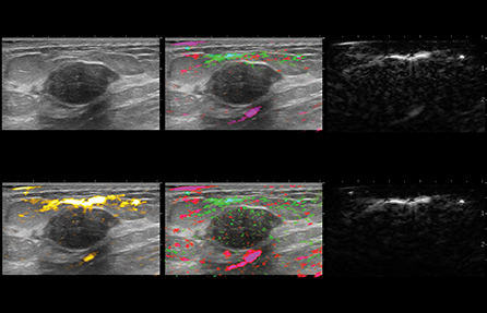
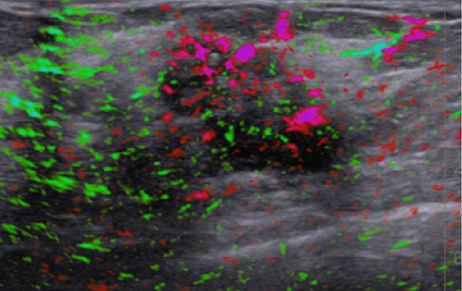
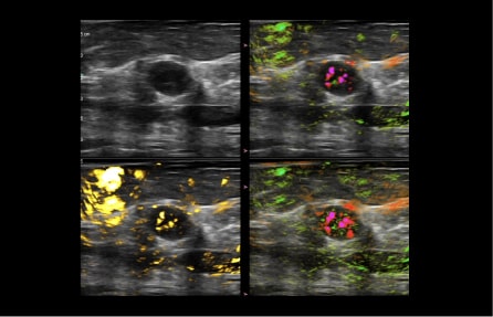
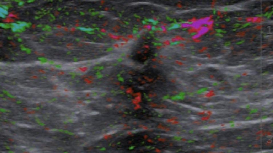
Signals from the sensors are analyzed and co-registered into images that present a real-time blood map of the lesion in easy to distinguish colors. OA image contrast is related to both blood volume and oxygenation status.
Blood within and around masses preferentially absorbs the light over normal tissue and becomes slightly heated. A transient thermoelastic expansion causes a tumor to emit a pressure or acoustic wave. This acoustic wave is then detected by sensors within the handheld, opto-acoustic (OA) probe as it is positioned over the indicated area of the breast.
A new view of cancer
In general, malignant masses are more vascular and deplete oxygen from the blood at a higher rate than do benign masses. The two wavelengths of laser light used in the Imagio® system facilitate imaging by showing the relative differences between oxygenated and deoxygenated blood.
Highly specific
With the Imagio® Breast Imaging System, clinicians have been able to identify and classify tumors as small as 3 millimeters, as well as visualize vascular structures smaller than one millimeter.

Our nationwide roadshow can bring Imagio® to your facility or you can start the conversation with a Seno rep today to get more details.